Best knee exercises for your healthy recovery after surgery.
Are knee exercises an important part of recovery after surgery?
The recovery period varies for every patient after knee replacement surgery. Most people, get 90% better by 6 weeks after the surgery. What you do in the recovery period really reflects the efficiency you are going to gain during that time. As already said, every patient is different and the needs for every patient to recover may be different. However total knee replacement exercises are a set of the most important elements of getting better after the surgery and no patient can avoid them. In order to regain strength and movement and to align your leg properly, you have to exercise from the first day after surgery. Regaining your ability to perform daily routine activities is a gradual process and depends upon what those activities are but you have to conform to these exercises done in the supervision of a good physiotherapist to even think about it.
How often do I have to exercise?
The nature of knee exercises after surgery will vary with time and will depend upon your progress. However, it is a general protocol that you get involved in these exercises in rounds of 25 minutes, at least 2 times a day. Two parts of your daily recovery routine after surgery are these sessions of exercises and walking for 30 minutes, 2 times a day.
Different Total knee replacement exercises at different stages of recovery?
For some weeks after surgery
Every set of exercises after surgery aims at a specific dimension of recovery. As the muscles in the leg after this surgery becomes weak and there is a threat of bad blood circulation, these early exercises aim at increasing the mobility of your knee joint, strengthening the muscles of your legs, and preventing the formation of blood clots which can be dangerous. Your orthopedic surgeon will recommend you start these knee exercises just after the surgery to set you on a path of early recovery. It is very obvious, these exercises will not feel pleasant and may even cause discomfort at first. However, being motivated can help you immensely to go through these recovery exercises. These exercises even help you to relieve the pain that comes after the surgery which can help you to avoid excessive use of painkillers.
Exercises for the frontal thigh (quadriceps)
This includes stretching the muscles of the thigh by laying down your leg to keep your knee straight. As you feel the tension in those muscles, you have to hold the position for 10 seconds and then release it. You have to perform 15 repetitions of this exercise, 2 times a day

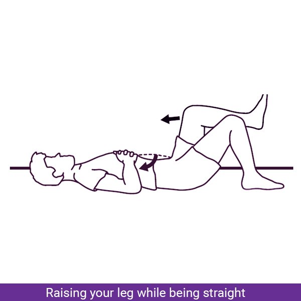
Raising your leg while being straight
This set of exercises also targets your thigh and aims to make your muscle strong again. What you have to do in this exercise is to stretch your thigh like mentioned above and raise your leg to the maximum height you can. You have to hold that position for about 10 seconds and then release.
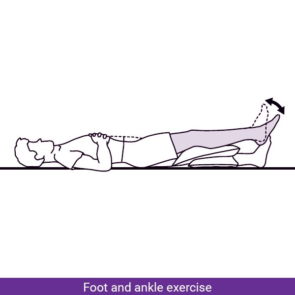
Foot and ankle exercise
This exercise includes pumping your ankle by moving your foot in an up-down motion continuously and slowly. This exercise will put tension in the area below your knee. You can do this exercise any time of the day as it is a very helpful and easy exercise to do.
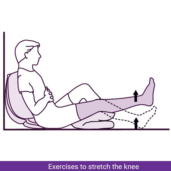
Exercises to stretch the knee
As there is generally a bend in your leg at the knee after surgery, it is important to do this set of total knee replacement exercises to get rid of that bend. For this exercise, you have to level up your foot by putting something like a rolled towel below the heel. You have to try to straighten your knee by stretching your leg in this position. At first, your knee may not be straight enough but as will progress it will recover to its normal alignment.
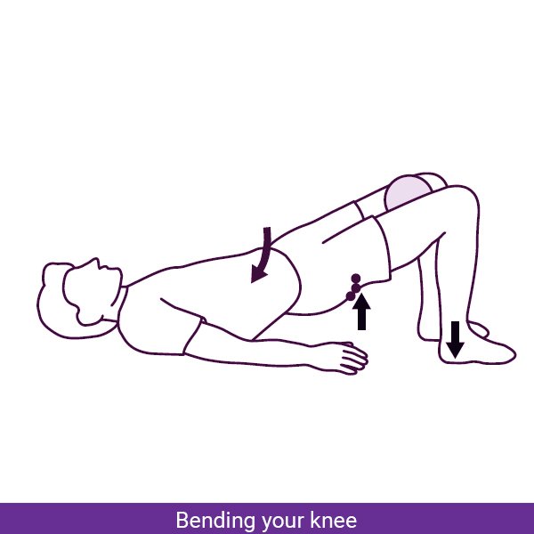
Bending your knee
This exercise is as important as straightening your knee in order to regain mobility. For this exercise, you have to lie down and slowly move your foot towards your hip while being on the bed surface. This will bend your knee and you have to hold the position of tension for 10 seconds. You can also do some variations of this exercise while sitting on a chair.
Knee exercises after a few weeks of recovery
For these exercises, you must have already gained some strength and the ability to move for some distance. As the damage to the knee because of your osteoarthritis and to the muscles around your knee because of the surgery makes the whole leg become weak, it takes dedication and time to recover with full strength. During this time you are recommended to do the following exercise.
Bending your knee while standing on a walker
For this exercise, you have to think of being in the gym and holding the position of maximum tension for at least 10 seconds
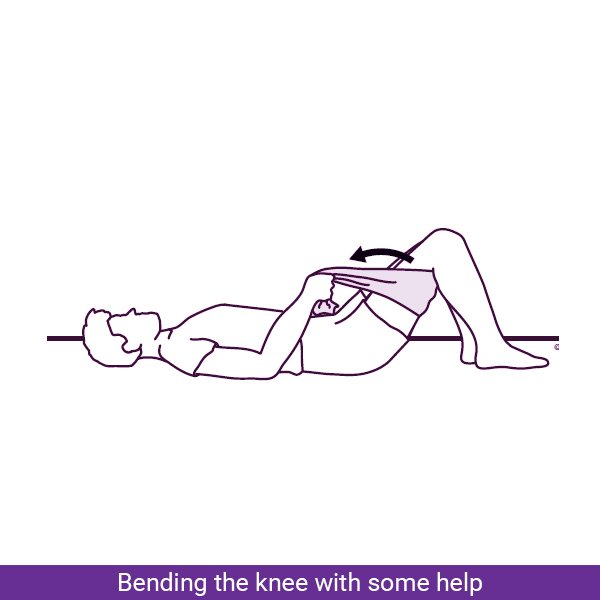
Bending the knee with some help
While lying down on a surface, you have to bend your knee while raising the foot for this exercise. You can use the help of some band or towel to get your foot close to the buttock.
Exercising with an elastic band
To strengthen your muscles more and more, you must push yourself to some level of discomfort. If you are recovered to the point when you are able to do the above exercises with ease, it is better to add some tension to them using an elastic band or even some weight.

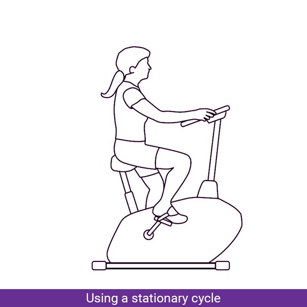
Using a stationary cycle
You may be able to do this cardio exercise after 5 to 6 weeks of recovery. Riding a stationary bike is one of the best exercises for your whole body to distribute weight and strength.
Conclusion
The total knee replacement exercises you do will certainly vary in nature. This is because knee replacement surgery can make your whole leg weak. You have to keep the progress of these knee exercises in check by walking for 30 minutes, 2 times a day, and even stepping up and down stairs. Your choice of a physiotherapist is as crucial as the choice of your orthopedic surgeon. We, at Om Physio Plus Nutrition, help you to get in touch with excellent practitioners who can make your recovery as efficient as possible.
To get in touch now, contact us at Om Physio Plus Nutrition